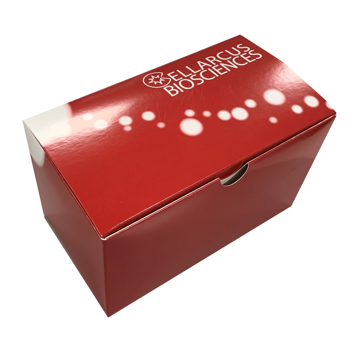
vFC™ VESICLE FLOW CYTOMETRY EV ANALYSIS ASSAY
FOR COUNTING AND SIZING VESICLES IN BIOFLUIDS, MEDIA, OR BUFFER
$450.00 – $2,100.00
Description
Sample Type: Suitable for characterization of extracellular vesicles directly from cell culture supernatant or biofluids such as plasma, urine, or CSF. Can also be used to analyze EVs separated from complex samples using various techniques. See kit manual for sample type specific assay considerations.
Species: Selected above. Human and mouse kits contain an antibody to label common EV surface proteins and an appropriate positive control. Although most reagents are not species specific, certain controls restrict the use of this kit to human samples.
Limits of Detection: vFC™ is suitable for the enumeration of vesicles down to 50nm from background. By following our staining protocols and using vTag™ monoclonal antibodies, surface antigen detection down to 10 molecules per vesicle is possible. Limit of detection is instrument specific. See product manual for additional information.
Cargo Measurement: vFC™ enables quantitative cargo measurement in a no-wash format using fluorescently labeled antibodies. We recommend vTag™ antibodies which perform optimally and have been appropriately titrated for vFC™.
Instrument Requirements: vFC™ can be run on sensitive, commercially available flow cytometers including the Beckman Coulter CytoFLEX®, the Luminex CellStream® and ImageStream®, and the Cytek® Aurora.
Sample Storage: Care should be taken to keep assay and storage conditions consistent and report all procedures. If vesicles cannot be analyzed fresh, we recommend storing at -80°C and aliquoting to avoid unnecessary freeze/thaw cycles.
Kit Storage: CAUTION! Multiple storage temperatures required. See product manual for instructions.
What are the limitations of the assay?
vFC™ assay (#CBS4) is intended for the analysis of vesicles from cell culture in supernatant or buffer. It is also useful for analyzing vesicles separated from biofluids. It can detect vesicles down to 50nm on currently available instrumentation. Limit of detection of surface markers per vesicle is approximately 10 molecules (depending on the instrument and the vTag™ antibody used). We have characterized limits for several instruments. Please contact us for instrument specific sensitivity assessments.
Why am I not seeing events from the Lipo100™ control sample?
There are several possible reasons for this issue. First, ensure that the instrument you’re using is suitable for vFC™. Next, make sure that the instrument has been properly set up and qualified. For instrument setup procedures see the instrument setup and configuration document under the datasheets and documentation tab. Finally, double check that samples are being properly diluted. Pay careful attention to details such as the plate being used for dilutions as certain plastics may bind vesicle and significantly impact EV counts. For additional support, chat with us by clicking on the icon at the bottom right of the window.
Why am I seeing high background events?
High number of background events can arise from multiple sources. Ensure proper dilution of samples and only run vesicles that are appropriate for this kit. Also, rarely carryover from samples may lead to background which is especially noticeable in blank samples. This is most commonly observed after running small beads. Run a daily clean cycle prior to continuing analysis. If issues continue, run beads diluted 1:10 in cleaning solution in the future.
CellStream® Protocols
CytoFLEX® Protocols
Cytek® Aurora Protocols
ImageStream® Protocols
Kit Overview
If this is your first time running vFC™, start with the appropriate instrument setup protocol for your flow cytometer and then complete protocol A before moving into analysis of your samples. This will ensure your instrument is performing well and will provide rough calibration of data to enable initial interpretation. Protocol A will also require vCal™ NANORAINBOW BEADS. Next, for all new samples, always run protocol 1. Protocol 1 provides essential controls to demonstrate EV specificity and singly EV resolution of the assay. Last, and for routine measurement of samples, run protocol 2 to determine the size distribution of EVs and to quantify EV concentration and copy number of various surface markers. Prior to publication and to ensure accuracy of the data from the assay run protocols B1-B3 and apply the instrument specific calibrations to your data as outlined in the protocols.
FCS Express and FCS Express Reader Analysis Layouts
CytoFLEX®
CellStream®
Cytek® Aurora
Sample Data
CytoFLEX®
CellStream®
- Lin Y, Anderson JD, Rahnama LMA, Gu S V., Knowlton AA. Exosomes in disease and regeneration: Biological functions, diagnostics, and beneficial effects.
Am J Physiol Heart Circ Physiol. 2020;319(6):H1162-H1180.
DOI: 10.1152/AJPHEART.00075.2020/ASSET/IMAGES/LARGE/AJ-AHRT200011F003.JPEG - Silva TF, Hutchins E, Zhao W, et al. Extracellular Vesicles heterogeneity through the lens of multiomics.
bioRxiv. Published online August 19, 2024:2024.08.14.607999.
DOI: 10.1101/2024.08.14.607999 - Oh CK, Dolatabadi N, Cieplak P, et al. S-Nitrosylation of p62 Inhibits Autophagic Flux to Promote α-Synuclein Secretion and Spread in Parkinson’s Disease and Lewy Body Dementia.
Journal of Neuroscience. 2022;42(14):3011-3024.
DOI: 10.1523/JNEUROSCI.1508-21.2022 - Chen C, Cai N, Niu Q, Tian Y, Hu Y, Yan X. Quantitative assessment of lipophilic membrane dye-based labelling of extracellular vesicles by nano-flow cytometry.
J Extracell Vesicles. 2023;12(8):12351.
DOI: 10.1002/JEV2.12351 - Woud WW, van der Pol E, Mul E, et al. An imaging flow cytometry-based methodology for the analysis of single extracellular vesicles in unprocessed human plasma.
Communications Biology 2022 5:1. 2022;5(1):1-14.
DOI: 10.1038/s42003-022-03569-5 - Crooks ET, Almanza F, D’Addabbo A, et al. Engineering well-expressed, V2-immunofocusing HIV-1 envelope glycoprotein membrane trimers for use in heterologous prime-boost vaccine regimens.
PLoS Pathog. 2021;17(10):e1009807.
DOI: 10.1371/JOURNAL.PPAT.1009807 - Schaubmayr W, Hochreiter B, Hunyadi-Gulyas E, et al. The Proteome of Extracellular Vesicles Released from Pulmonary Microvascular Endothelium Reveals Impact of Oxygen Conditions on Biotrauma.
Int J Mol Sci. 2024;25(4):2415.
DOI: 10.3390/IJMS25042415/S1 - Sako Y, Sato-Kaneko F, Shukla NM, et al. Identification of a Novel Small Molecule That Enhances the Release of Extracellular Vesicles with Immunostimulatory Potency via Induction of Calcium Influx.
ACS Chem Biol. 2023;18(4):982-993.
DOI: 10.1021/ACSCHEMBIO.3C00134/ASSET/IMAGES/LARGE/CB3C00134_0007.JPEG - Thom SR, Bennett M, Banham ND, et al. Elevations of Extracellular Vesicles and Inflammatory Biomarkers in Closed Circuit SCUBA Divers.
International Journal of Molecular Sciences 2023, Vol 24, Page 5969. 2023;24(6):5969.
DOI: 10.3390/IJMS24065969 - Yan W, Cao M, Ruan X, et al. Cancer-cell-secreted miR-122 suppresses O-GlcNAcylation to promote skeletal muscle proteolysis.
Nature Cell Biology 2022 24:5. 2022;24(5):793-804.
DOI: 10.1038/s41556-022-00893-0 - Shpigelman J, Lao FS, Yao S, et al. Generation and Application of a Reporter Cell Line for the Quantitative Screen of Extracellular Vesicle Release.
Front Pharmacol. 2021;12:668609.
DOI: 10.3389/FPHAR.2021.668609/BIBTEX - Sandau US, Duggan E, Shi X, et al. Methamphetamine use alters human plasma extracellular vesicles and their microRNA cargo: An exploratory study.
J Extracell Vesicles. 2020;10(1):e12028.
DOI: 10.1002/JEV2.12028 - Tekkatte C, Lindsay SA, Duggan E, et al. Identification of optimal conditions for human placental explant culture and extracellular vesicle release.
iScience. 2023;26(10).
DOI: 10.1016/j.isci.2023.108046 - Nguyen DB, Tran HT, Kaestner L, Bernhardt I. The Relation Between Extracellular Vesicles Released From Red Blood Cells, Their Cargo, and the Clearance by Macrophages.
Front Physiol. 2022;13:783260.
DOI: 10.3389/FPHYS.2022.783260/BIBTEX - Choi W, Park DJ, Dorschner RA, Nakatsutsumi K, Yi M, Eliceiri BP. CDK1-loaded extracellular vesicles promote cell cycle to reverse impaired wound healing in diabetic obese mice.
Molecular Therapy. 2025;0(0).
DOI: 10.1016/J.YMTHE.2025.01.039 - Park DJ, Choi W, Sayeed S, et al. Defining the activity of pro-reparative extracellular vesicles in wound healing based on miRNA payloads and cell type-specific lineage mapping.
Molecular Therapy. 2024;32(9):3059-3079.
DOI: 10.1016/J.YMTHE.2024.02.019/ASSET/508B2B9E-A0D2-4E80-91DC-11E6902DAD10/MAIN.ASSETS/GR3.JPG - Welsh JA, Van Der Pol E, Arkesteijn GJA, et al. MIFlowCyt-EV: a framework for standardized reporting of extracellular vesicle flow cytometry experiments.
J Extracell Vesicles. 2020;9(1):1713526.
DOI: 10.1080/20013078.2020.1713526 - Beckner ME, Conkright WR, Mi Q, et al. Neuroendocrine, inflammatory, and extracellular vesicle responses during the Navy Special Warfare Screener Selection Course.
Physiol Genomics. 2022;54(8):283-295.
DOI: 10.1152/PHYSIOLGENOMICS.00184.2021/ASSET/IMAGES/LARGE/PHYSIOLGENOMICS.00184.2021_F007.JPEG - Vieira E, Nolan J, Maslanik T, et al. Liquid Brain Biopsy in Late-Life Depression and Alzheimer’s Disease Using Circulating Brain-Derived Exosomes.
Biol Psychiatry. 2022;91(9):S40.
DOI: 10.1016/j.biopsych.2022.02.117 - Park DJ, Duggan E, Ho K, et al. Serpin-loaded extracellular vesicles promote tissue repair in a mouse model of impaired wound healing.
J Nanobiotechnology. 2022;20(1):1-17.
DOI: 10.1186/S12951-022-01656-7/FIGURES/6 - Patel A, Patel S, Patel P, Mandlik D, Patel K, Tanavde V. Salivary Exosomal miRNA-1307-5p Predicts Disease Aggressiveness and Poor Prognosis in Oral Squamous Cell Carcinoma Patients.
International Journal of Molecular Sciences 2022, Vol 23, Page 10639. 2022;23(18):10639.
DOI: 10.3390/IJMS231810639 - Sandau US, McFarland TJ, Smith SJ, Galasko DR, Quinn JF, Saugstad JA. Differential Effects of APOE Genotype on MicroRNA Cargo of Cerebrospinal Fluid Extracellular Vesicles in Females With Alzheimer’s Disease Compared to Males.
Front Cell Dev Biol. 2022;10:864022.
DOI: 10.3389/FCELL.2022.864022/BIBTEX - Nakatsutsumi K, Choi W, Johnston W, et al. Lung contusion complicated by pneumonia worsens lung injury via the inflammatory effect of alveolar small extracellular vesicles on macrophages and epithelial cells.
Journal of Trauma and Acute Care Surgery. Published online 2024.
DOI: 10.1097/TA.0000000000004499 - Costantini TW, Park DJ, Johnston W, et al. A heterogenous population of extracellular vesicles mobilize to the alveoli postinjury.
Journal of Trauma and Acute Care Surgery. 2024;96(3):371-377.
DOI: 10.1097/TA.0000000000004176 - Gao Y, Jablonska A, Chu C, Walczak P, Janowski M. Mesenchymal Stem Cells Do Not Lose Direct Labels Including Iron Oxide Nanoparticles and DFO-89Zr Chelates through Secretion of Extracellular Vesicles.
Membranes 2021, Vol 11, Page 484. 2021;11(7):484.
DOI: 10.3390/MEMBRANES11070484 - Conkright WR, Beckner ME, Sterczala AJ, et al. Resistance exercise differentially alters extracellular vesicle size and subpopulation characteristics in healthy men and women: an observational cohort study.
Physiol Genomics. 2022;54(9):350-359.
DOI: 10.1152/PHYSIOLGENOMICS.00171.2021 - Schaubmayr W, Hochreiter B, Hunyadi-Gulyas E, et al. The Proteome of Extracellular Vesicles Released from Pulmonary Microvascular Endothelium Reveals Impact of Oxygen Conditions on Biotrauma.
Int J Mol Sci. 2024;25(4):2415.
DOI: 10.3390/IJMS25042415/S1 - Kim J, Xu S, Jung SR, et al. Comparison of EV characterization by commercial high-sensitivity flow cytometers and a custom single-molecule flow cytometer.
J Extracell Vesicles. 2024;13(8).
DOI: 10.1002/JEV2.12498 - Park DJ, Choi W, Sayeed S, et al. Defining the activity of pro-reparative extracellular vesicles in wound healing based on miRNA payloads and cell type-specific lineage mapping.
Molecular Therapy. 2024;32(9):3059-3079.
DOI: 10.1016/J.YMTHE.2024.02.019/ASSET/508B2B9E-A0D2-4E80-91DC-11E6902DAD10/MAIN.ASSETS/GR3.JPG - Narayanan P, Shen L, Curtis BR, et al. Investigation into the Mechanism(s) That Leads to Platelet Decreases in Cynomolgus Monkeys During Administration of ISIS 104838, a 2’-MOE-Modified Antisense Oligonucleotide.
Toxicol Sci. 2018;164(2):613-626.
DOI: 10.1093/TOXSCI/KFY119
Additional information
| Size | 96 Wells, 5 x 96 Wells |
|---|---|
| EV Marker (Antibody) ⓘ | Anti-Human TS Cocktail (CD9, CD63, CD81) [PE], Anti-Human TS Cocktail (CD9, CD63, CD81) [PE-Cy7], Anti-Mouse TS Cocktail (CD9, CD63, CD81) [PE], Anti-Mouse TS Cocktail (CD9, CD63, CD81) [PE-Cy7], Other, None |
FOR COUNTING AND SIZING VESICLES IN BIOFLUIDS, MEDIA, OR BUFFER” Cancel reply


Reviews
There are no reviews yet.